
Study reveals how positive emotions trigger sudden muscle weakness in narcolepsy

A newly published study is providing fresh insight into cataplexy, a symptom of narcolepsy that can cause sudden muscle weakness or collapse. By examining the activity of specific brain neurons and receptors, Auburn University College of Veterinary Medicine researcher Dr. Daniel Kroeger helped discover that oxytocin is a key driver of these events, identifying a potential new target for future therapies.
Kroeger, a neuroscience professor, studies sleep and the ways it impacts memory, emotions and health conditions like Alzehimer’s disease. Guided by the college’s One Health mission, his research spans animal and human health, seeking insights that improve outcomes across species.
Before coming to Auburn, Kroeger embarked on the study with his colleague at Harvard University, Dr. Carrie Mahoney. He played a key role in incorporating optogenetics into the project, drawing on expertise he developed during a postdoctoral fellowship at Stanford University. The technique uses pulses of light to activate targeted neurons, allowing researchers to precisely investigate their role in complex biological processes.
“Quite often in the brain, you have neurons that do one thing intermingled with neurons that do the opposite,” Kroeger said. “If you were to advance a stimulating electrode in the middle of them and turn on the juice, you would activate both. Now, with optogenetics, we can use light flashes to activate only the brain cells that we want to activate and leave their neighbors untouched.”
Using both optogenetics and chemogenetics — another technique used to control the activity of specific cells — the research team targeted specific neurons to assess their role during a cataplexy episode.
Scientists have long known that narcolepsy is linked to the loss of orexin neurons, specialized brain cells that help stabilize wakefulness, but much less is known about the broader network of brain cells involved in the disorder. In a healthy person, orexin neurons are active during waking periods and inactive during sleep — a bit like a light switch. Orexin communicates with sleep and wake neurons, telling them when and when not to inhibit the other.
People with narcolepsy are missing a significant portion of orexin neurons. Those without more than 80% of orexin neurons experience severe narcolepsy often accompanied by cataplexy when exposed to triggers like laughter and strong social interactions. For the mice in the study, triggers included eating chocolate and being reunited with siblings after 20 minutes of separation.
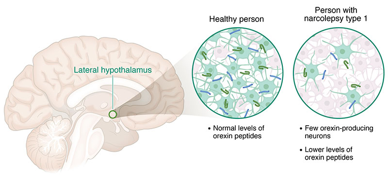
Image from “Mignot and Yanagisawa win Lasker prize for discovery of the role of orexins in wake, sleep, and narcolepsy,” The Journal of Clinical Investigation (2026).
“So, the question was, what is the signal that is produced by happiness that leads to cataplexy?” Kroeger said.
The project grew out of two key observations. Previous studies showed that the central nucleus of the amygdala — a major processing center in the brain for emotions — was an integral player in cataplexy. Researchers also knew that a hormone called oxytocin is released in the brain during positive social interactions and rewarding experiences. Mahoney and Kroeger set out to investigate whether there was a connection between the oxytocin released during positive experiences and cataplexy episodes.
The team started by measuring oxytocin levels in the central nucleus of the amygdala in narcoleptic mice. They discovered that oxytocin increased just before cataplexy triggered by eating chocolate or strong social interactions. Using optogenetics, they activated neurons in the hypothalamus that produce oxytocin and found that activation triggered cataplexy. Next, the team stimulated oxytocin receptors in the amygdala, which also triggered cataplexy. Those two outcomes confirmed their hypothesis that happiness-induced oxytocin release in the amygdala directly triggers cataplexy.
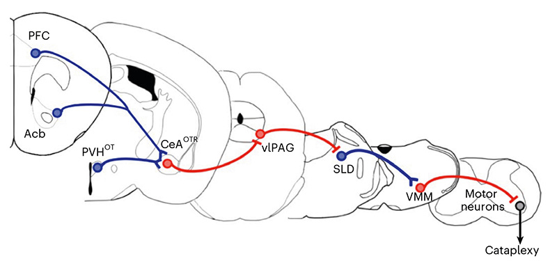
Image from “Oxytocin promotes socially triggered cataplexy,” Nature Neuroscience (2026).
From there, the researchers were able to chart the downstream pathways, discovering that the oxytocin-responsive neurons in the central nucleus of the amygdala cause cataplexy by inhibiting the neurons in the brainstem that usually keep muscles from going limp during waking periods.
While there are pharmaceuticals in development to reduce cataplexy by addressing the lack of orexin in the brain, Kroeger said this discovery could help researchers target cataplexy from a different angle.
“This helps our understanding of how the brain works,” Kroeger said. “Understanding the pathways and knowing that cataplexy is oxytocin-driven could lead to the design of some kind of antibody that prevents oxytocin from docking on to specific cells and triggering cataplexy.”
Faculty Research